|
Dental caries is a disease that damages
tooth structures, resulting
in what is commonly called tooth decay or cavities, which are holes in the teeth. This damage first affects the hard tissues
of the teeth (enamel, dentin and cementum). As the destruction
progresses, these tissues begin to break down, which can eventually lead to holes in the teeth. If left untreated, the disease can lead to pain, tooth loss, infection, and, in severe
cases, death.There is a long
history of dental caries: over a million years ago, hominids such as Australopithecus suffered from
cavities. However, the incidence of cavities was
very low well into the Paleolithic and Mesolithic periods.
The largest increases in the prevalence of caries have been associated with dietary changes. Today, caries remains one of
the most common diseases throughout the world.
There are numerous ways to classify dental caries. Although the presentation may differ, the risk
factors and development among distinct types of caries remain largely similar. Initially, it may appear as a small chalky
area but eventually develop into a large, brown cavitation. Though sometimes caries may be seen directly, radiographs are
frequently needed to inspect less visible areas of teeth and to judge the extent of destruction.
Tooth decay is caused by certain types of acid-producing bacteria (specifically
Lactobacillus species, Streptococcus
mutans, and Actinomyces
species) which cause damage in the presence of fermentable carbohydrates such as sucrose, fructose, and glucose. The resulting
high levels of acidity from lactic acid in the mouth affect
teeth because a tooth's special mineral content causes
it to be sensitive to low pH. Specifically,
a tooth (which is primarily mineral in content) is in a constant state of back-and-forth demineralization and remineralization between the tooth
and surrounding saliva. When the pH at
the surface of the tooth drops below 5.5, demineralization proceeds faster than remineralization (i.e. there is a net loss
of mineral structure on the tooth's surface). This results in the ensuing decay. Depending on the extent of tooth destruction,
various treatments can be used to restore teeth to proper
form, function, and
aesthetics, but there is
no known method to regenerate large amounts
of tooth structure. Instead, dental health organizations advocate preventive and prophylactic measures, such as regular oral hygiene and
dietary modifications, to avoid dental caries.
|
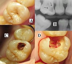
| A. Pit Caries B.Radiographic view of Cl II caries C. Cl II cavity D. After caries excavation |
|